Diagnostic Radiology
Diagnostic Radiology
The use of X-rays for diagnosis is licensed by the Federal Authority for Nuclear Regulation (FANAR).
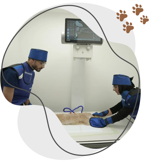
X-ray services at a Welfare veterinary clinic are a fundamental diagnostic tool used to quickly assess an animal’s internal structures, especially bones and certain organs, without surgery.
These services use low doses of radiation to produce images that help veterinarians identify issues such as fractures, joint problems, lung diseases, and swallowed foreign objects. X-rays are also useful for evaluating the size and shape of organs like the heart and for checking dental health in some cases.
The procedure is typically fast and non-invasive. Animals may need to be gently restrained or lightly sedated to ensure clear images and minimize stress.
Overall, X-ray services provide valuable, immediate insights that support accurate diagnosis and effective treatment planning for a wide range of conditions.
Advanced X-ray imaging of animal fractures
Advanced X-ray imaging for animal fractures goes beyond standard radiography to provide highly detailed views that improve diagnosis and treatment planning. It is especially useful for complex, subtle, or hard-to-locate fractures.
Key features include:
- Digital radiography (DR): Produces high-resolution images that can be enhanced (zoomed, adjusted for contrast) to better visualize small or complicated fractures.
- Computed radiography (CR): Offers improved image quality over traditional film and allows for digital storage and analysis.
- Multiple view positioning: Taking images from different angles helps identify fracture type, alignment, and displacement more accurately.
- Contrast studies (when needed): Highlight specific structures if fractures involve joints or surrounding tissues.
These techniques allow veterinarians to:
- Detect hairline or stress fractures
- Assess fracture severity and alignment
- Plan surgical or non-surgical treatment
- Monitor healing over time
Overall, advanced X-ray imaging provides precise, fast, and minimally invasive evaluation, leading to better outcomes in fracture management.
We Are the Best
Benefits of X-rays for lung injuries in pets
X-rays are one of the most valuable first-line tools for evaluating lung injuries in pets because they provide fast, non-invasive insight into the chest. Key benefits include:
Rapid assessment: X-rays can quickly reveal life-threatening conditions such as lung contusions (bruising), pneumothorax (air in the chest cavity), or fluid buildup, allowing for immediate treatment decisions.
Clear visualization of the lungs and chest: They help identify abnormalities like inflammation, bleeding, collapsed lung areas, or masses that may not be visible externally.
Detection of trauma-related damage: After accidents or falls, X-rays can show rib fractures, diaphragm issues, or associated chest injuries affecting breathing.
Guidance for treatment planning: Findings help veterinarians decide whether oxygen therapy, surgery, or further imaging is needed.
Monitoring recovery: Repeated X-rays allow vets to track healing progress and ensure conditions like fluid or air accumulation are resolving.
Overall, X-rays provide a quick, accessible, and reliable way to diagnose and manage lung injuries in pets, improving the chances of timely and effective care.

Visit Us On @Instagram